
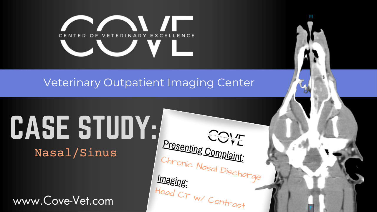
Nasal/Sinus Disease
From chronic sneezing to nasal discharge or facial swelling, our CT imaging provides a clear, detailed view to guide treatment or surgical planning.
Click Here to View Case Study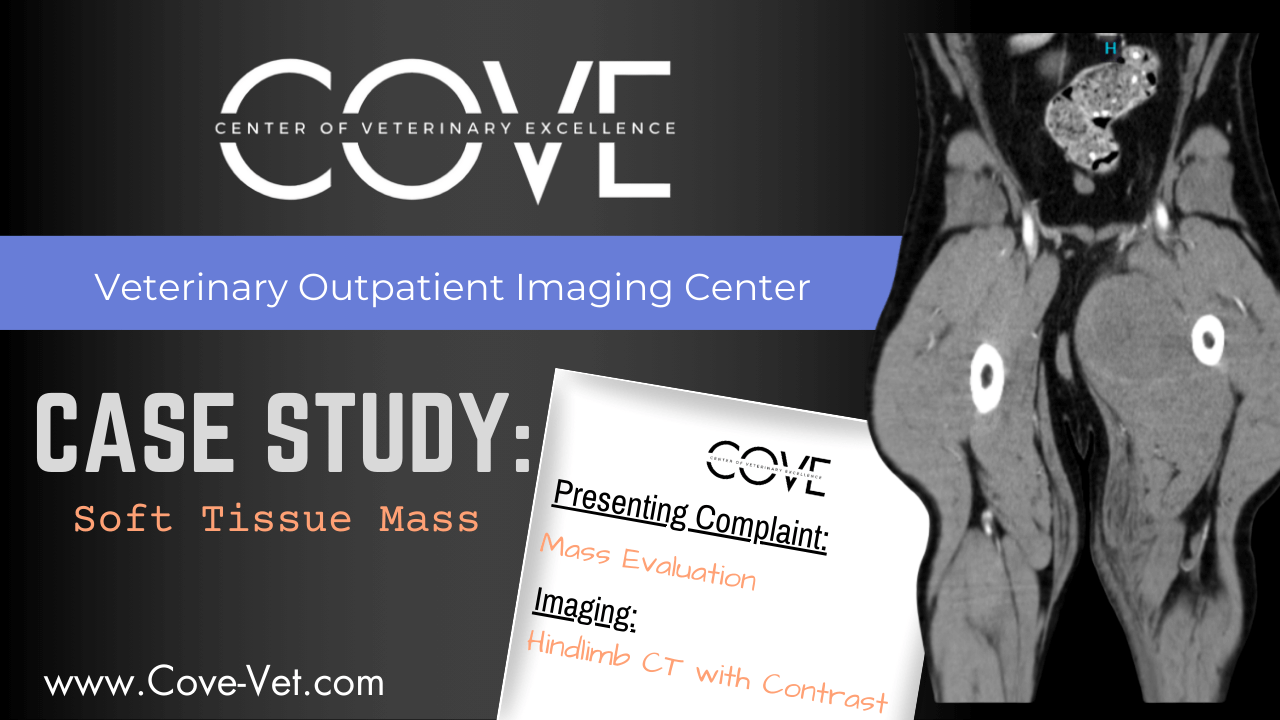
Soft Tissue Mass Evaluation
When a mass is detected, time matters. Our CT imaging helps distinguish soft tissue tumors from surrounding structures, assess invasiveness, and support surgical or oncologic planning with clarity and speed.
Click Here to View Case Study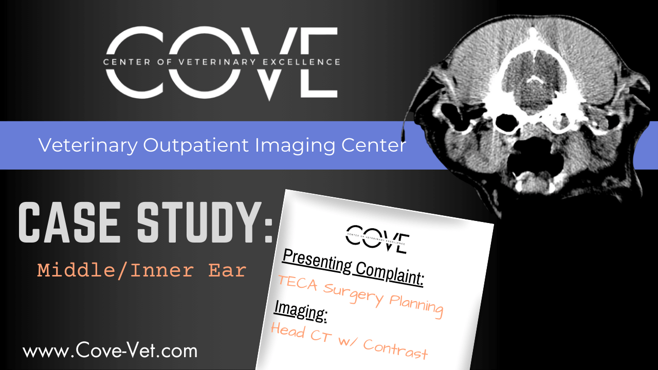
Middle/Inner Ear Evaluation
If your patient shows signs of head tilt, balance issues, or persistent ear infections, we offer precise imaging to assess inner ear structures and guide appropriate care.
Click Here to View Case Study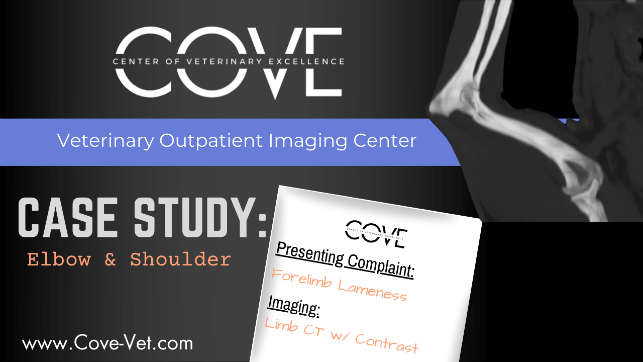
Elbow & Shoulder Evaluation
CT imaging offers superior detail for diagnosing elbow dysplasia, fragmented coronoid process, osteochondritis dissecans (OCD), and other joint abnormalities—helping guide treatment for forelimb lameness.
Click Here to View Case Study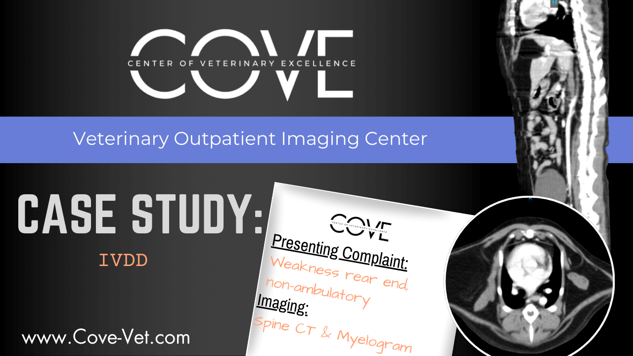
Intervertebral Disc Disease (IVDD)
Our advanced spinal CT imaging helps localize disc herniation, compression, or degeneration—providing critical information for timely neurologic treatment or surgical planning.
Click Here to View Case Study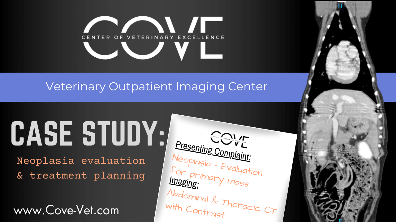
Neoplasia Evaluation & Treatment Planning
CT imaging provides essential detail for characterizing masses, assessing local invasion or metastasis, and guiding surgical or oncologic treatment strategies.
Click Here to View Case Study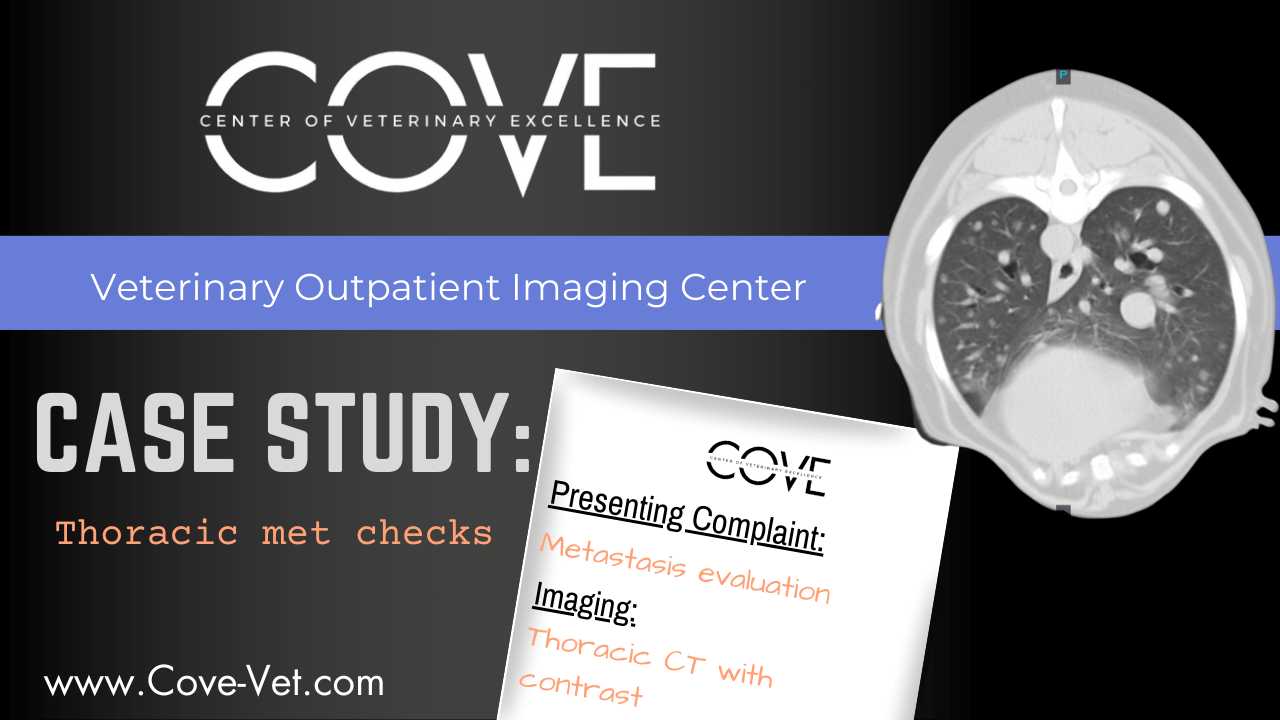
Thoracic Met Checks
Quick, clear scans help evaluate for metastasis or primary thoracic tumors—critical in planning next steps in treatment.
Click Here to View Case Study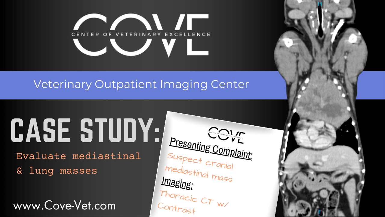
Evaluate Mediastinal & Lung Masses
CT imaging offers a detailed view of thoracic structures to localize masses, differentiate tissue types, and support surgical or oncologic planning with clarity.
Click Here to View Case Study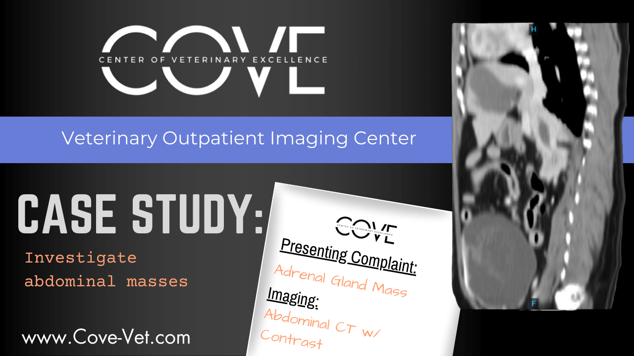
Investigate Abdominal Masses
Investigate and localize masses, assess for metastasis, and prepare for surgery with clear anatomical detail.
Click Here to View Case Study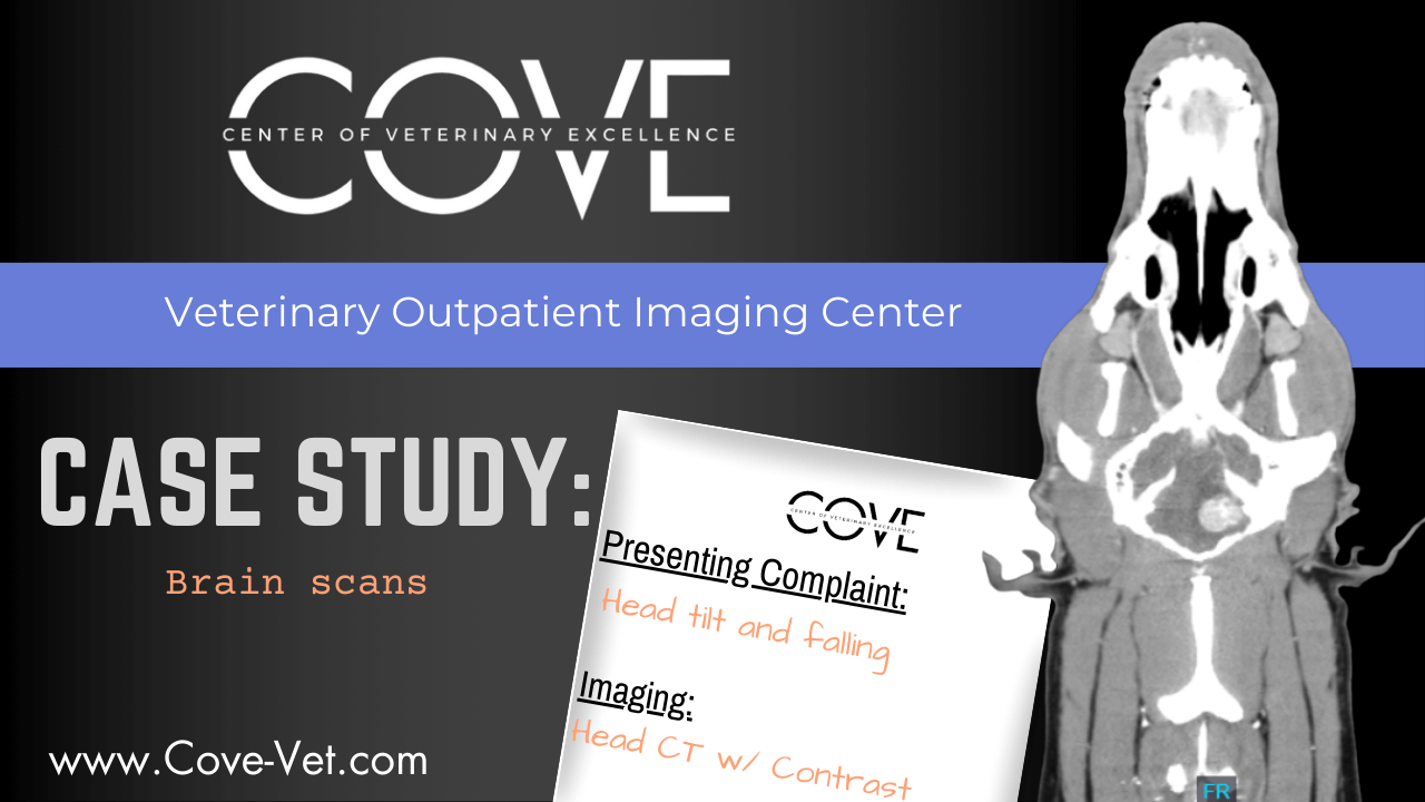
Brain Imaging
Our brain scans support neurologic evaluations with high-definition imaging to detect abnormalities, or tumors.
Click Here to View Case Study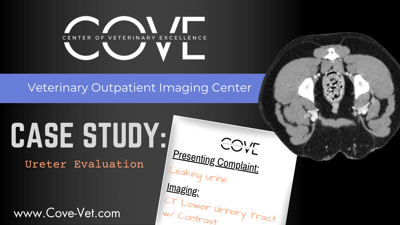
Ureter Evaluation
CT imaging allows precise identification of ureteral obstructions, and ectopic ureters, supporting treatment decisions.
Click Here to View Case Study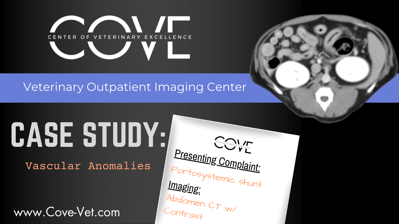
Vascular Anomalies
CT imaging allows for precise identification of vascular malformations, aneurysms, or shunts, aiding in accurate diagnosis and treatment planning for complex circulatory issues.
Click Here to View Case Study


