Waukesha, WI: (262) 668-7878 |   Farmington Hills, MI: (947) 500-2683

ECHOCARDIOGRAMS  

Advanced
Cardiac Care

Comprehensive cardiac evaluation goes beyond imaging alone. Standardized protocols, detailed measurements, and board-certified cardiology interpretation work together to provide accurate diagnoses and informed treatment recommendations.

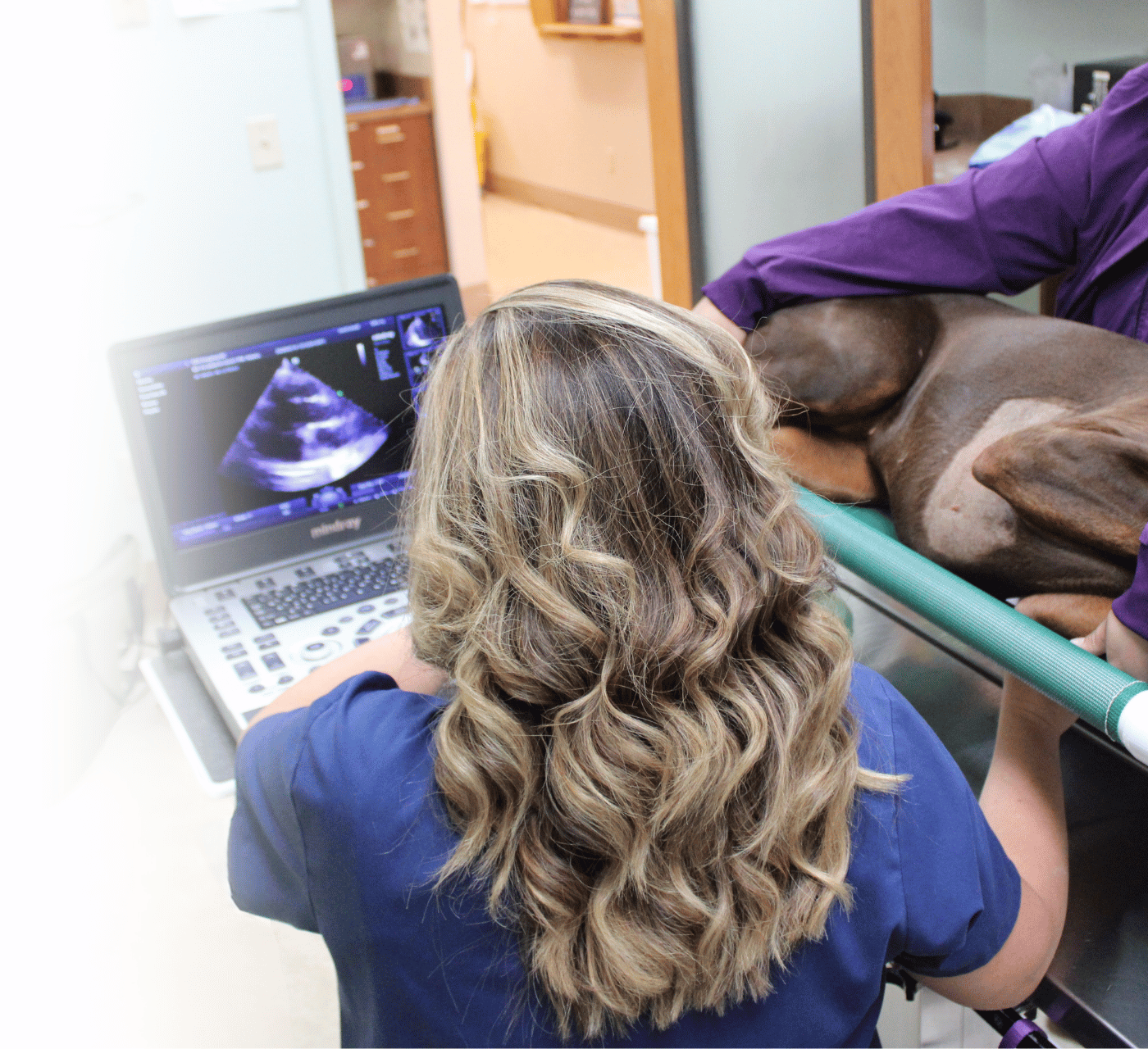
 

Veterinary echocardiogram performed to evaluate heart structure and function

ECHOCARDIOGRAMS

Advanced
Cardiac Care

Comprehensive cardiac evaluation goes beyond imaging alone. Standardized protocols, detailed measurements, and board-certified cardiology interpretation work together to provide accurate diagnoses and informed treatment recommendations.
 

Echo Service
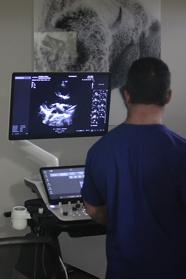
Ultrasound imaging of a canine heart during comprehensive echocardiographic examination
When are Echocardiograms Recommended?
COMMON APPLICATIONS
 
Echocardiography is recommended when cardiac disease is suspected or when additional information is needed to evaluate heart function, monitor disease progression, and support treatment planning.
What's Included During an Echocardiogram?
COMPREHENSIVE EVALUATION
 
Every echocardiogram follows standardized imaging protocols to evaluate heart anatomy, blood flow, and overall cardiac function. Multiple imaging views and advanced diagnostic techniques provide a thorough assessment, helping guide diagnosis, treatment recommendations, and ongoing patient care.
Canine patient positioned on a cardiac table with phased array probe during ultrasound exam
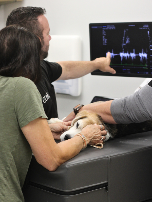
Veterinary technician and pet owner gently holding dog during echocardiogram at the COVE
Veterinary sonographer obtaining necessary images and measurements for board-certified cardiologist review
Echocardiogram in progress for canine patient
BEYOND THE ECHOCARDIOGRAM
 
ELECTROCARDIOGRAM (EKG/ECG)
 
An electrocardiogram (EKG/ECG) is obtained either before or during the echocardiogram to assess heart rate and rhythm
and to detect for any arrhythmias.
 
 
THORACIC RADIOGRAPH REVIEW
 
When available, thoracic radiographs may be reviewed by the cardiologist to assess cardiac size, vessel size, and findings related to congestive heart failure. Thoracic radiographs should be obtained by the primary veterinary team and provided as part of the referral information to support the cardiac evaluation.
 
 
Please note: Cardiologist review of thoracic radiographs is limited to findings related to cardiac structure and function. Other abnormalities that may be present on the radiographs will not be evaluated or commented on. If a complete interpretation of thoracic radiographs is desired, review by a board-certified radiologist is recommended.
Patient and Pet Owner Echocardiogram
Comfort Designed for You & Your Pet
PERSONALIZED PATIENT CARE
 
Whenever appropriate, pet owners may be invited to remain with their pet during portions of the examination. Our team is committed to creating a calm, supportive environment while ensuring every study is performed safely and effectively.
Complete Examination 
Includes:
 
  • Long-axis
  • Short-axis

Comprehensive long-axis and short-axis imaging to evaluate cardiac anatomy, chamber size, and valve function.

Includes:
 
  • 4-Chamber
  • 5-Chamber

Captures detailed four- and five-chamber views to assess cardiac structure, blood flow, and ventricular function from the heart's apex.

Includes:
 
  • Mitral Valve
  • LA/Ao
  • Papillary Muscles
  • Chordae Tendineae
  • Aortic Valve
  • Pulmonary Artery

Detailed evaluation of key cardiac structures to identify abnormalities affecting heart function, valve performance, and blood flow.

Includes:
 
  • Subcostal Views
  • Cranial Abdominal Views (When Indicated)

Additional imaging is obtained when clinically appropriate to provide further diagnostic information and support a complete cardiac evaluation.

Includes:
 
  • Blood Flow Evaluation
  • Velocity Measurements 

Measures the direction and speed of blood flow through the heart and major vessels, helping identify abnormal circulation and valve disorders.

Includes:
 
  • Chamber Measurements
  • Wall Motion Assessment 

Provides precise measurements of cardiac chamber size, wall thickness, and heart motion to evaluate overall cardiac function.

Includes:
 
  • Color Blood Flow Mapping
  • Valve Assessment 

Uses color mapping to visualize the direction and pattern of blood flow, making it easier to identify turbulence, leaks, and abnormal valve function.

Includes:
 
  • Targeted Flow Analysis
  • Velocity Measurements

Measures blood flow velocity at specific locations within the heart and blood vessels to evaluate normal and abnormal cardiac function.

Includes:
 
  • High-Velocity Flow Analysis
  • Pressure Gradient Assessment

Evaluates high-velocity blood flow across narrowed or abnormal heart valves, helping assess the severity of cardiac disease.

Board-Certified Expertise
Every echocardiogram performed at The COVE is reviewed by board-certified cardiologist committed to delivering accurate interpretations, timely reporting, and collaborative support. Meet our specialists, review case studies, or refer a patient to get started.

MEET THE SPECIALISTS

Learn about the board-certified veterinary specialists behind every CT interpretation and the expertise they bring to every case.

VIEW CASE STUDIES

Explore real-world CT cases and discover how advanced imaging supports accurate diagnoses and informed treatment decisions.

REFER A PATIENT

Give us a call or fill out our online form to schedule an appointment.

Waukesha, WI: (262) 668-7878
Farmington Hills, MI: (947) 500-2683